
Atomic Force Microscopy
Guarantor
Mohamed Bel Moubarik, Assistant Professor
Instrument status:
Operational 01/04/2024
Equipment placement:
Riad 8 first floor, Physics low temperature Area
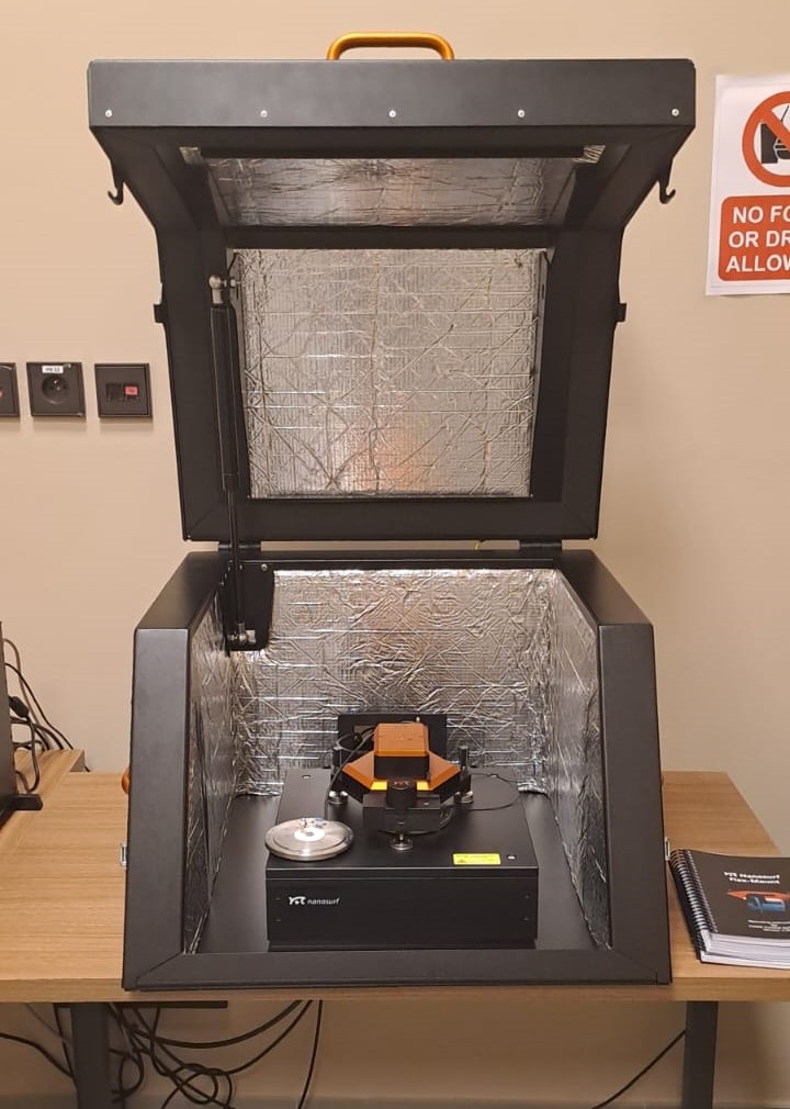
The Nanosurf FlexAFM system is an atomic force microscope that can measure the topography and several other properties of a sample with nanometer resolution. These measurements are performed, displayed, and evaluated using the Nanosurf C3000 control software.
The Nanosurf FlexAFM combined with the C3000 controller is a fully-fledged research system that can be used to perform stand-alone AFM measurements or be integrated into inverted microscope systems for combined microscopic (fluorescence, phase contrast, brightfield) and AFM data (topography, elasticity, force, etc.).
The available measurements modes are the following: Static (Contact Mode), Dynamic (Tapping Mode), Kelvin probe force microscopy (KPFM), piezoresponse force microscopy (PFM), magnetic force microscopy (MFM) and STM mode for the atomic level microscopy.
- Specifications and features:
| Scan Head Specifications | FlexAFM 100 μm |
| Sample size | Unlimited without sample stage; 100 mm on sample stage |
| Maximum Petri dish height (fluid level in dish) | 9 mm (6 mm) |
| Manual approach range | 30 mm |
| Manual approach range | 1.1 mm |
| Maximum scan range | 100 μm (1) |
| Maximum Z-range | 10 μm (2) |
| Drive Z-resolution | 0.152 nm (3) |
| Drive XY-resolution | 1.525 nm (3) |
| XY-linearity mean error typ. 0.1% | typ. 0.1% |
| XY-flatness at maximum scan range typ. 5 nm typ. 1 nm | typ. 5 nm |
| Z-measurement noise level(RMS, Static Mode in air) | typ. 0.3 nm |
| Z-measurement noise level (RMS, Dynamic Mode in air) | typ. 0.16 nm |
| Scan head dimensions | 143 × 158 × 53 mm |
| Scan head weight | 1.25 kg |
(1) Manufacturing tolerances are ± 5%
(2) Manufacturing tolerances are ± 10%
(3) Calculated by dividing the maximum range by 16 bits
- FlexAFM Scan Head Features
| General design | Tripod stand-alone, flexure-based electromagnetically actuated XY-scanner, piezo based Z-scanner |
| Cantilever alignment | Automatic alignment for cantilevers with alignment grooves. Manual laser adjustment possible for special cantilevers. |
| Laser adjustment | No laser adjustment required upon immersion of cantilever into liquid because of SureAlign™ laser optics |
| Laser adjustment | Available |
| Laser adjustment | Top and side view in air and liquid |
| Visual magnification | Top: 13× / Side: 10× |
| Sample illumination | White axial illumination for top and side view. Transmission illumination with illuminated sample holder. |
| Cantilever holder | Cleanable and with replaceable cantilever spring |
| Operating modes | Static Force, Dynamic Force, Phase Contrast, MFM / EFM, Spreading Resistance, Force Modulation, Lateral Force, Kelvin Probe Force, Scanning Thermal, Multiple Spectroscopy modes, Lithography and Manipulation modes |
- AFM Options
| Operation modes | explanations |
|
Static (Contact Mode) |
Static mode, or contact mode, is the original and simplest mode to operate an AFM. In this mode, the probe is in continuous contact with the surface while the probe raster scans the sample. The most common configuration of static mode is to operate it in constant force or deflection feedback mode. In this mode, the cantilever deflection is the feedback parameter. |
| Dynamic (Tapping Mode) |
Dynamic force mode refers to a collection of AFM modes in which the cantilever oscillates at a high frequency at or close to resonance. A specific kind of dynamic mode, referred to as amplitude modulation mode (AM-AFM) is the most common AFM imaging mode. In AM AFM, the amplitude of oscillation is the feedback parameter; other dynamic modes have different parameters for the feedback loop such as frequency (frequency modulation) or phase (phase modulation). |
| Kelvin probe force microscopy |
Kelvin probe force microscopy (KPFM) is an extension of the dynamic force mode. Using KPFM, images can be recorded that contain information on the local work function or local contact potential difference between tip and sample. |
| PFM | This static mode-based method is geared towards the study of ferroelectric or piezoelectric materials, which are materials that respond mechanically to the application of an electric field. This mode measures topography simultaneously with mechanical response of the material when an electric voltage is applied with a conductive AFM tip. A sharp conductive AFM tip is brought into contact with the sample and an AC voltage is applied between the tip and sample. The amplitude gives information on the piezoelectric tensor of the material and the phase provides information on the polarization direction. |
| MFM | Magnetic force microscopy (MFM) is a phase imaging mode that uses atomic force microscope cantilevers with a thin magnetic coating in order to probe the magnetic field between a sample and a magnetized tip. This method is commonly used to image any materials with heterogeneous magnetic properties such as magnetic-based hard disk drives. It can be operated in single, interlaced and dual scan line modes. Any of these modes require optimization of the height above the sample at which the magnetic force microscopy image is collected. |
| STM | The NaioSTM is a scanning tunneling microscope that brings together scan head and controller in a single instrument for even simpler installation, maximized ease of use, and straightforward transportability. The setup is robust against vibrations and can be used to achieve atomic resolution on HOPG in standard classroom situations. |