
Research
About IAP
Equipment
About
Users Committee
Equipment
Raman Spectroscopy
Terahertz Imaging
Atomic Force Microscopy
Synthesis Area
Magneto Transport Spectroscopy
Femto-laser
Fees and Chemical Information
User Access
Reservation/Booking
Semi-Conductors Physics
Terahertz Photonics
Nuclear, Particle Physics, and Cosmology
Space Science and Technology
OCP Track
Publications
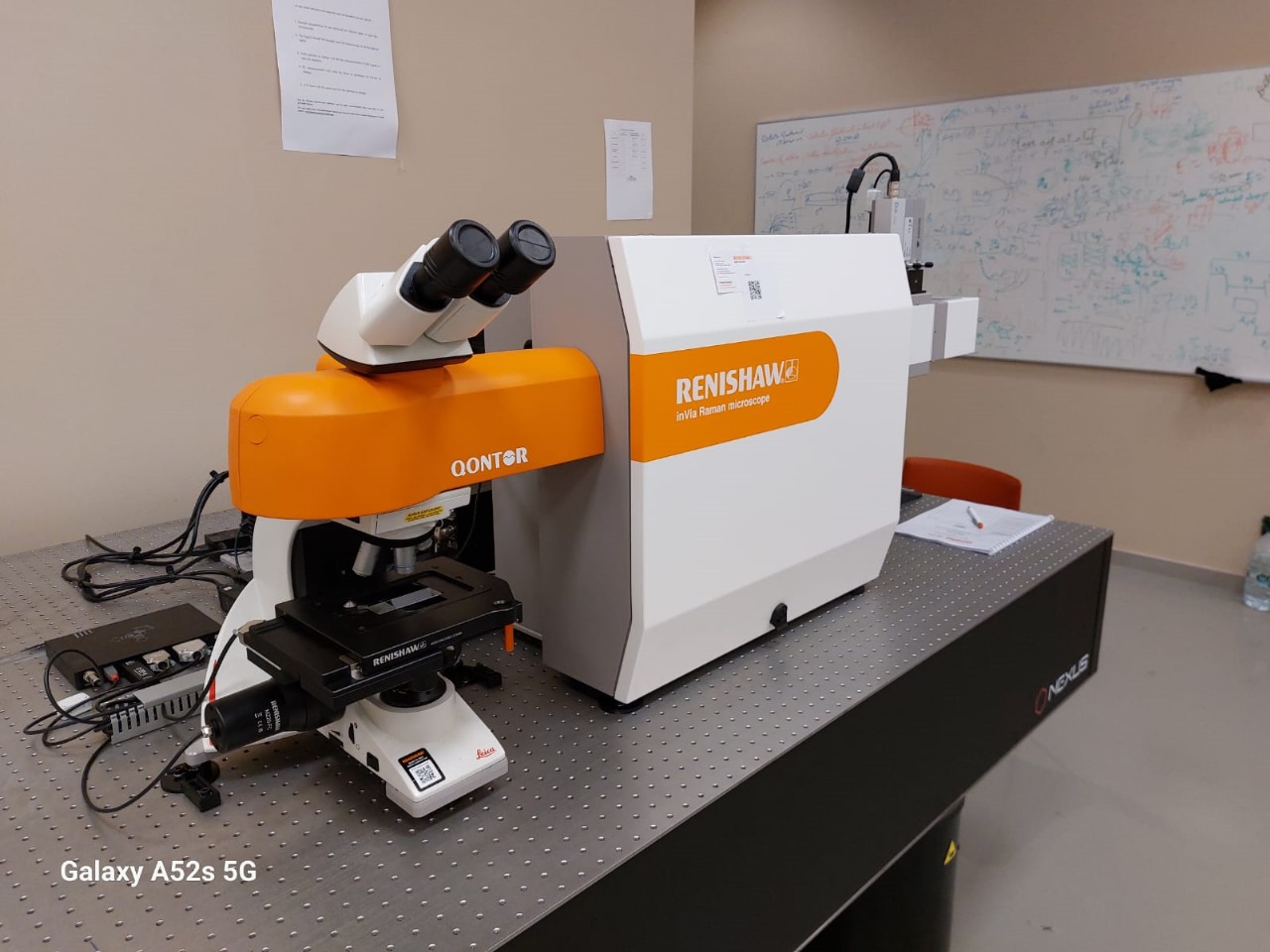
The inVia confocal Raman spectroscopy
Guarantor:
Mohammed Amlieh, Education fellow
Instrument status:
Operational 27/03/2024 11:07
Equipment placement:
Riad 8 first floor, Physics low temperature Area,
The inVia confocal Raman imaging system offers outstanding performance in speed, sensitivity and resolution, as well as reliable results for even the most challenging experiments. It comprises a research-grade microscope coupled to a high-performance Raman spectrometer. Hence it can produce both rich, detailed, chemical images and highly specific data from discrete points.
 Specification:
Renishaw inVia confocal spectrometer for Raman, PL, and multispectral imaging at 355, 532 and 785 nm
Spectrometer Features:
| Equipment | Description |
| Stigmatic spectrograph | Single stage, 250 mm focal length, very bright (throughput > 30 % in the spectrograph |
| Beam expander | Motorised and optimised for each exciter for working at the diffraction limit with all working configurations |
| Mirrors | Independent and optimised for each exciter for maximum performance |
| Input slit | Motorised, software-controlled |
| CCD detector | Renishaw centre’s NIR/UV deep depletion Peltier cooled (-70 C) 1-inch 1024 pixels (26 microns/pixel). Requires no water or liquid nitrogen |
| Neutral density filters | 16 power levels (from 0.00005 to 100%) available directly from the software |
Basic Microscope Features:
Research grade optical microscope with objectives:
- 10x (NA 0.25, WD 17.6mm)
- 20x (NA0.40, WD 1.15mm)
- 50x (NA 0.50, WD 8.20 mm)
Motorized stage travelling range:
- 112 x 76 mm in X, Y
- 25 mm in Z with 8 nm resolution
Step size:
- 50 nm in X, Y
- 8 nm in Z
Excitation Lasers:
| Wavelength (nm) | Power range (mW) | Laser blocking filter () |
| 355 | 10 | 150 |
| 532 | 50 | 100 |
| 785 | 300 | 80 |